


Medical Tests
Imaging tests are critical diagnostic tools, allowing physicians to see what's going on inside your body. These tests use various forms of energy (x-rays, sound waves, radioactive particles, or magnetic fields) to create amazing images of your tissues and organs. While they tend to be remarkably safe, imaging tests that utilize ionizing radiation are cause for some concern. The benefits that these tests provide must be balanced against the risks associated with radiation exposure.
Studies have found a clear link between radiation and cancer. Most of these studies have looked at people exposed to very high doses of radiation, however, and the risk from low-level radiation exposure is more difficult to determine. Nevertheless, the impact of medical testing radiation appears to be substantial: Some experts estimate that up to 2% of all cancer cases are caused by it.
The radiation exposure from the average diagnostic x-ray increases cancer risk only slightly, likely on the order of hundredths to thousandths of one percent. But the effects of radiation exposure add up over a lifetime. The more imaging tests you undergo, the higher your risk of cancer. For that reason, imaging tests that use radiation should be used judiciously and performed only when they're clearly indicated.
X-rays
X-rays use electromagnetic radiation to create images of certain areas inside the body and can be used to diagnose a variety of medical conditions. The amount of radiation used is quite small. A single chest x-ray exposes the patient to about 0.1 mSv, which is about the radiation dose people receive naturally over the course of 10 days. A mammogram (an x-ray of the breast) exposes a woman to 0.7 mSv, or about the amount of radiation a person would expect to get in about 3 months.
To make x-rays as safe as possible, a lead apron should be placed over parts of the body that are not being studied, especially areas that are particularly sensitive to radiation such as the reproductive organs. Pregnant women should get x-rays only when absolutely necessary since radiation exposure can cause birth defects.
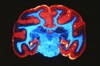
Computerized Tomography Scans
Computerized tomography scans, also called CT scans or "Cat" scans, use special x-ray equipment and computers to create pictures of the body. During the procedure, an x-ray unit rotates around the body, creating three dimensional images. The amount of radiation received during a typical CT scan is larger than the radiation received from other methods of x-ray imaging.
Some medical imaging facilities offer "whole body CT scanning/screening" as a preventive healthcare measure to individuals with no symptoms of illness. There is very little evidence that whole body CT scans are effective screening tests. For a person who is not experiencing symptoms or who has no specific health concern, whole body CT scanning is likely to cause more harm than good. Public health agencies and numerous national medical and professional societies do not typically recommend CT scanning for such purposes.
Nuclear Scans
Nuclear scans are a bit like an inside out x-ray. Instead of directing radiation through a patient's body, they introduce radioactive materials into the body and then measure the energy that is given off by them. The radioactive materials are typically injected into a vein, swallowed or inhaled as a gas. Different types of materials are attracted to different tissues and organs in the body and eventually accumulate there. Once they do, a special type of camera detects the radioactivity that they give off and records the information.
Common nuclear medicine procedures include thyroid studies, brain scans, bone scans, lung scans, cardiac stress tests, and liver and gallbladder procedures.
Not all imaging tests utilize ionizing radiation.
Ultrasound
Ultrasound machines DO NOT use radiation. Instead, they emit high-frequency sound waves that travel through the body. The waves pass right through certain tissues and are deflected by others. Waves that bounce back (or 'echos') are recorded by the ultrasound machine and converted into images.
MRI
MRI scanners also DO NOT use radiation. They create images using magnetic fields and radio frequency waves.
In some cases, they can be used in place of one of the tests that does. But different tests provide doctors with different types of images and different information about the body. Ultrasounds, for example, are great tests if gallstones are the problem; they're a poor choice, however, if the problem is a brain tumor. In many cases, there simply isn't a good substitute and a test that uses radiation is the way to go.
Although no one wants to be exposed to radiation unnecessarily, fear of exposure should never stop someone who really needs an imaging study from getting it.
Links
- Radiation and Medical X-rays - U.S. Environmental Protection Agency
- Medical X-ray Imaging - U.S. Food and Drug Administration
- X-Rays, Pregnancy and You - U.S. Food and Drug Administration
- Computed Tomography (CT) - U.S. Food and Drug Administration
- Computed Tomography (CT) Scans and Cancer - National Cancer Institute
- Full-Body CT Scans - What You Need to Know - U.S. Food and Drug Administration